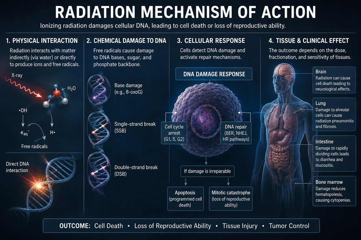
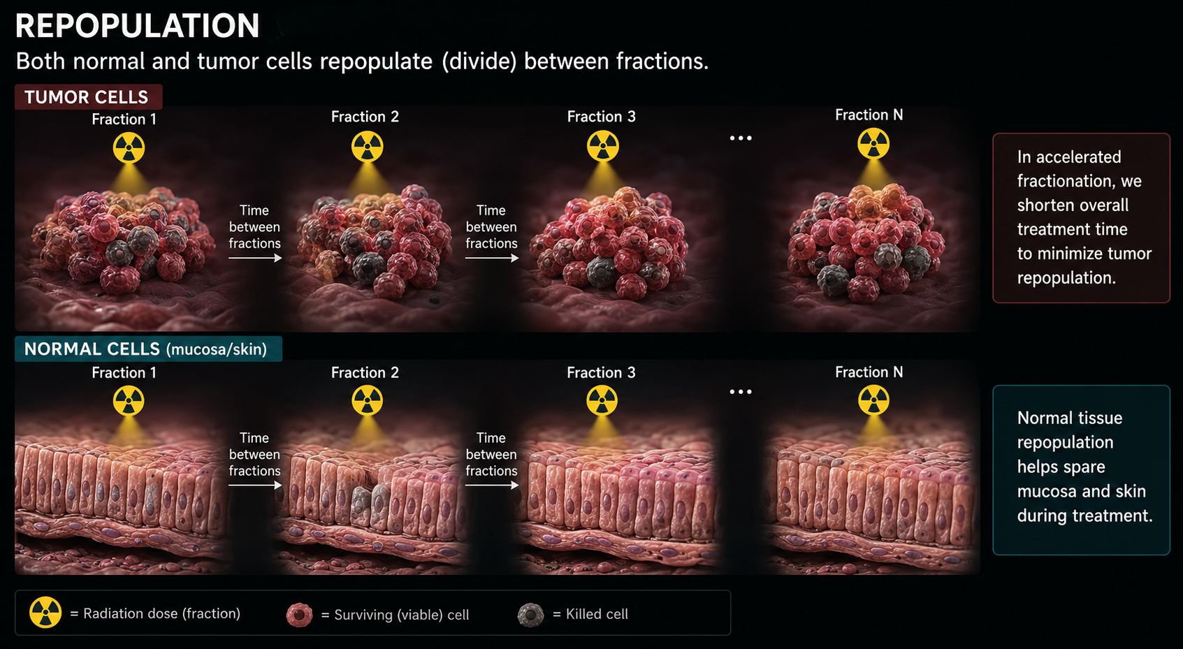
🎯 Modality 6

Radiotherapy

Radiotherapy uses high doses of ionizing radiation to damage cancer cell DNA, preventing them from dividing. It treats ~50% of all cancer patients at some point in their care — either with curative or palliative intent.

50%
Cancer Patients Treated
6–18
MV Photon Energy (LINAC)
Gray
Unit of Absorbed Dose
1896
First Radiation Treatment

The 4 R's of Radiobiology

Understanding why we use fractionated (divided) doses over multiple days requires understanding how radiation affects cells — both tumor cells and normal tissue. The four R's explain how fraction timing improves tumor control while protecting normal tissues.

How Radiation Kills Cancer Cells
Radiation mechanism of action infographic showing physical interaction, DNA damage, cellular response, and tissue clinical effects
Repair radiobiology infographic comparing normal cells that repair sublethal DNA damage between fractions with tumor cells that accumulate damage and die

1. Repair

Normal tissue recovers between fractions

The image shows daily radiation doses injuring both normal and tumor cells, but normal cells repair sublethal DNA damage during the hours to days between fractions. Tumor cells repair less effectively, so damage accumulates across treatments.

Clinical point: fractionation exploits the repair advantage of normal tissue, improving the therapeutic ratio.

Redistribution reassortment radiobiology infographic showing tumor cells shifting through G1, S, G2, and M cell-cycle phases after repeated fractions

2. Redistribution / Reassortment

Cells shift into more radiosensitive phases

The image maps tumor cells across the cell cycle before treatment and after repeated daily fractions. Late S-phase cells are more resistant, while mitotic and G2 cells are more sensitive; repeated fractions gradually catch more cells in vulnerable phases.

Clinical point: spacing fractions gives tumor populations time to reassort into phases where radiation is more effective.

Reoxygenation radiobiology infographic showing hypoxic inner tumor cells becoming oxygen rich and more radiosensitive before the next fraction

3. Reoxygenation

Hypoxic tumor cells become easier to kill

The image contrasts an oxygen-poor tumor core during the first fraction with improved oxygen delivery before the next fraction. As outer tumor cells die and blood flow reaches deeper cells, previously hypoxic cells become reoxygenated and more radiosensitive.

Clinical point: oxygen fixes radiation-induced DNA damage, so reoxygenation makes later fractions more lethal to the tumor.

Repopulation radiobiology infographic showing tumor cells and normal mucosa or skin cells dividing between radiation fractions

4. Repopulation

Cells divide during the treatment course

The image shows both tumor cells and normal mucosa or skin cells dividing between fractions. Normal tissue repopulation helps healing, but tumor repopulation can reduce control if the overall treatment course is stretched too long.

Clinical point: accelerated schedules may be used to limit tumor repopulation while still allowing normal tissue recovery.

Linear Accelerator (LINAC)

Labeled components of a modern medical linear accelerator used for external beam radiotherapy.

LINAC components infographic labeling electron gun, accelerator, bending magnet, X-ray target, primary collimator, multi-leaf collimator, gantry, treatment couch, and control system

LINAC Components

The linear accelerator (LINAC) is the standard machine for external beam radiotherapy. The labeled image identifies the major treatment and control components.

🎯

A modern LINAC delivers high-energy X-rays (6–18 MV) or electrons (4–22 MeV) to the tumor from multiple gantry angles, with the tumor at the isocenter. Sophisticated shaping of the beam (MLC, IMRT, VMAT) maximizes dose to tumor while minimizing dose to surrounding normal tissue.

Treatment Techniques

Radiotherapy has evolved from simple opposed beams to highly conformal, motion-adapted delivery — dramatically improving the therapeutic ratio.

Photorealistic 3D conformal radiotherapy infographic showing fixed shaped beams converging on a tumor target.

3D-CRT — 3D Conformal RT

CT-based treatment planning with beams shaped by collimators/MLCs to conform to the 3D shape of the target. The modern baseline for external beam RT. Multiple fixed beam angles from 3D planning CT.

Photorealistic IMRT infographic showing MLC leaf modulation and varied beamlet intensity shaping dose around organs at risk.

IMRT — Intensity Modulated RT

MLC leaves move during delivery, creating non-uniform (modulated) fluence within each beam. Allows "dose painting" — high dose to tumor, steep dose gradient to organs at risk. Step-and-shoot or sliding-window techniques.

Photorealistic VMAT infographic showing a LINAC rotating in an arc while dose conforms around a tumor target.

VMAT — Volumetric Modulated Arc Therapy

Gantry rotates continuously while MLC positions, dose rate, and gantry speed all vary simultaneously. Highly conformal delivery in 1–3 arcs. Often faster than IMRT. RapidArc® (Varian) is a proprietary VMAT system.

Photorealistic SBRT and SABR infographic showing narrow high-dose beams focused on a small body tumor with immobilization.

SBRT / SABR — Stereotactic Body RT

Very high dose per fraction (>5 Gy/fx) in 3–5 fractions. Precise targeting of small tumors (lung, liver, spine, prostate). Requires sub-millimeter accuracy and immobilization. Requires IGRT and motion management.

Photorealistic stereotactic radiosurgery infographic showing micro-arcs converging on a small intracranial lesion.

SRS — Stereotactic Radiosurgery

Single high-dose treatment (15–25 Gy) for intracranial targets. GammaKnife® uses ~200 Co-60 sources; Linac-based SRS uses multiple micro-arcs. Used for brain mets, AVM, acoustic neuroma, trigeminal neuralgia.

Photorealistic brachytherapy infographic showing an afterloader source path and steep close-range dose falloff.

Brachytherapy

Radioactive sources placed inside or adjacent to tumor. HDR (high dose rate) — Ir-192 afterloader moves through catheters. LDR (low dose rate) — permanent seeds (I-125, Pd-103) or temporary implants. Used in prostate, cervix, breast, skin cancers.

Photorealistic proton therapy infographic showing a beam stopping at the Bragg peak inside a tumor with minimal exit dose.

Proton Therapy

Protons deposit most energy at the Bragg peak — abrupt stop at calculated depth with minimal exit dose. Ideal for pediatric tumors and structures adjacent to critical organs. Requires large cyclotron/synchrotron facility.

Photorealistic IGRT infographic showing onboard imaging, registration overlays, couch shifts, and target alignment before treatment.

IGRT — Image-Guided RT

Daily imaging (kV, CBCT, ExacTrac, MRI-LINAC) to verify patient positioning and target location before each fraction. Accounts for inter-fraction variation in organ position, tumor shrinkage, and weight loss.

Treatment Planning Process

Radiotherapy requires meticulous planning to ensure the right dose is delivered to the right place, every fraction.

Photorealistic CT simulation setup with patient immobilization in a radiation oncology CT room.
1

Simulation (CT Sim)

Patient positioned in treatment position (reproducible immobilization devices: masks, cradles, boards). Planning CT acquired. MRI/PET may be fused for target delineation.

Photorealistic radiation oncology contouring workstation with CT slices and colored target outlines.
2

Target & OAR Contouring (Delineation)

Radiation oncologist delineates: GTV (gross tumor), CTV (clinical target — includes microscopic spread margin), PTV (planning target — CTV + setup uncertainty margin). Organs at risk (OARs) also outlined: spinal cord, lung, bowel, bladder, heart, etc.

Photorealistic radiotherapy dose prescription and fractionation review at a clinical planning workstation.
3

Dose Prescription & Fractionation

Prescription: total dose (Gray) / number of fractions / dose per fraction / OAR dose constraints. Standard fractionation: ~2 Gy/day, 5 days/week. Hypofractionation: larger doses, fewer fractions (e.g., 3–5 Gy/fx for prostate). Hyperfractionation: smaller fractions, twice daily.

Photorealistic medical physicist using a treatment planning system with beam geometry and dose maps.
4

Treatment Planning (TPS)

Medical physicist designs beam arrangement (angles, energies, weights), MLC shapes, and optimization objectives in the Treatment Planning System (TPS) (e.g., Eclipse, RayStation, Pinnacle). Dose-volume histograms (DVH) assess plan quality. Iterative optimization to meet PTV coverage and OAR constraints.

Photorealistic radiotherapy patient-specific quality assurance phantom measurement at a LINAC.
5

Plan Approval & QA

Radiation oncologist approves the plan. Medical physicist performs independent monitor unit (MU) calculation verification and patient-specific QA (e.g., IMRT QA with phantom). Patient-specific QA verifies dose delivery before first treatment.

Photorealistic radiation therapists delivering treatment with a patient positioned on a modern LINAC couch.
6

Treatment Delivery (RTT Role)

Radiation Therapists (RTTs) set up and treat patients daily. IGRT imaging (CBCT) performed each fraction. RTTs monitor patient tolerance, document treatment, and maintain immobilization accuracy throughout the treatment course.

Clinical Applications

Radiotherapy is used in all major cancer types, either as primary treatment or combined with surgery and chemotherapy.

Gamma Knife radiosurgery unit used for stereotactic treatment of brain and central nervous system lesions.

🧠 Brain & CNS

Whole-brain RT, focal boost post-surgery, SRS for brain mets. Concurrent temozolomide + RT for GBM (Stupp protocol). Craniospinal irradiation for medulloblastoma.

Modern medical linear accelerator used for precision external beam radiotherapy, including thoracic tumors.

🫁 Thorax

Curative SBRT for early-stage inoperable lung cancer. Concurrent chemoradiotherapy for locally advanced NSCLC. Mesothelioma palliative RT. Prophylactic cranial irradiation in SCLC.

Cherenkov light image from a patient undergoing whole breast irradiation with a 6 MV radiotherapy beam.

🏋️ Breast

Post-lumpectomy whole-breast RT (WBI) or partial-breast irradiation (APBI). Post-mastectomy RT (PMRT) for high-risk disease. Modern hypofractionation: 40 Gy/15 fractions replaces 50 Gy/25 fractions.

Clinical brachytherapy procedure setup representing internal radiotherapy used for pelvic cancers.

🏥 Pelvis

Prostate: definitive EBRT or brachytherapy. Cervix: concurrent cisplatin + EBRT + brachytherapy (curative). Rectal: neoadjuvant CRT pre-surgery. Bladder: bladder-preservation trimodality therapy.

Thermoplastic immobilization mask used to reproduce head and neck positioning during radiotherapy.

🦷 H&N / ENT

Nasopharyngeal, oropharyngeal, laryngeal cancers. IMRT for salivary gland sparing. Concurrent platinum-based chemoradiotherapy. Organ preservation for larynx cancer.

Three-dimensional CT rendering of hip bone metastases, a common target for palliative radiotherapy.

💊 Palliative RT

Bone metastases (8 Gy single fraction for pain relief). Brain metastases (WBRT or SRS). Spinal cord compression (urgent). Obstruction/bleeding palliation. SBRT for oligometastatic disease.

👩‍⚕️

The Radiation Therapist's (RTT's) role: RTTs are the primary clinicians responsible for daily treatment delivery. They position patients with reproducibility, operate the LINAC, perform and review IGRT imaging, manage immobilization devices, monitor and document patient toxicity, and communicate with the multidisciplinary team throughout the treatment course. It is a technically demanding, patient-focused clinical role at the intersection of physics, technology, and care.

Well Done!

You've explored all 6 imaging modalities plus the history of radiology. Review any section, or go back to compare modalities side by side.

Back to Home