CT vs MRI: Which Imaging Modality and When— A Practical Guide
The Question Every Student Asks
"Should this patient get a CT or an MRI?" It's the most common imaging decision in modern medicine, and the answer is rarely straightforward. Both are cross-sectional modalities that revolutionized diagnostic imaging — but they work on completely different physical principles and excel at different things.
TL;DR — The 5-Second Rule
CT = fast, great for bone, acute bleeding, and trauma. Uses ionizing radiation.
MRI = detailed soft tissue contrast, no radiation. Slower, more expensive, more contraindications.
How They Work — The Physics Simplified
CT (Computed Tomography)
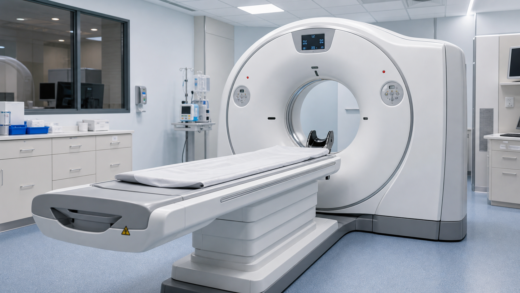
A CT scanner rotates an X-ray tube and detector array around the patient, acquiring hundreds of projections from different angles. A computer reconstructs these into cross-sectional (axial) slices using filtered back projection or iterative reconstruction. Each voxel in the image represents a Hounsfield unit — a measure of X-ray attenuation relative to water (0 HU). Air = -1000 HU, water = 0 HU, bone = +400 to +1000 HU, metal = +2000+ HU.
Key traits: Excellent spatial resolution, fast acquisition (seconds), excellent for bone and acute hemorrhage. Exposes the patient to ionizing radiation (effective doses typically 2-10 mSv for a CT, compared to 0.1 mSv for a chest X-ray).
MRI (Magnetic Resonance Imaging)
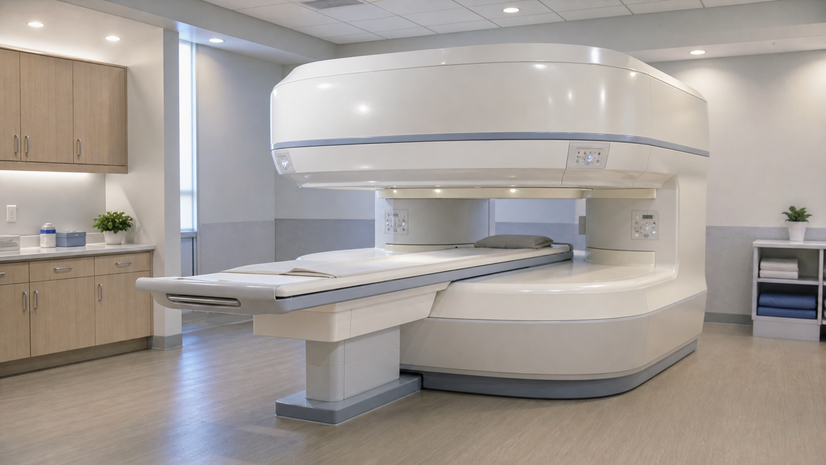
MRI uses powerful magnetic fields and radio waves — no ionizing radiation involved. (typically 1.5T or 3T) and radiofrequency pulses to excite hydrogen protons in the body. When the RF pulse stops, protons relax back to their equilibrium state, emitting signals that are spatially encoded by gradient coils and reconstructed into images. The key parameters — T1 relaxation, T2 relaxation, proton density, and diffusion — produce different tissue contrasts.
Key traits: Superior soft tissue contrast, no ionizing radiation, multiplanar capability without repositioning the patient. But: slow (20-60+ minutes), expensive, loud, and contraindicated for many implants.
Head-to-Head: Where Each Excels
| Clinical Question | Best Modality | Why |
|---|---|---|
| Acute head trauma | CT | Fast, excellent for skull fractures and acute hemorrhage (blood is hyperdense on CT) |
| Suspected stroke (acute) | CT | CT perfusion + CT angiography rule out hemorrhage and show salvageable tissue. "Time is brain." |
| Brain tumor characterization | MRI | Superior soft tissue contrast, gadolinium enhancement patterns, MR spectroscopy |
| Multiple sclerosis | MRI | FLAIR sequences show demyelinating plaques exquisitely. CT is nearly useless for MS. |
| Spinal trauma / fracture | CT | Bone detail is superior. CT is the first-line for C-spine clearance in trauma. |
| Spinal cord compression | MRI | Visualizes the cord, disc herniation, epidural abscess, and metastases. CT myelography is an alternative. |
| Pulmonary embolism | CT | CT pulmonary angiography (CTPA) is the gold standard. Fast, widely available. |
| Liver lesion characterization | MRI | Multi-phase contrast MRI with hepatobiliary agents is superior. CT is a good screening tool. |
| Kidney stones | CT | Non-contrast CT (KUB CT) has >95% sensitivity. Stones are radiopaque. MRI can't see stones well. |
| Knee internal derangement | MRI | Meniscal tears, ACL/PCL injuries, and cartilage defects. CT arthrography is a second-line option. |
| Shoulder rotator cuff | MRI | Excellent soft tissue contrast. Ultrasound is also good. CT is not appropriate. |
Contraindications — When You Can't Use Each
CT Contraindications
- Pregnancy — Ionizing radiation to the fetus is a risk. Minimize or defer. Use ultrasound or MRI when possible.
- Iodinated contrast allergy — Pre-medication protocol or switch to non-contrast study or MRI.
- Renal impairment — Iodinated contrast can cause contrast-induced nephropathy. Check eGFR.
- Thyroid storm risk — Iodinated contrast can trigger thyrotoxicosis in patients with hyperthyroidism.
MRI Contraindications (Absolute)
- Cardiac pacemakers (non-MRI-conditional) — The magnetic field can disrupt pacing. MRI-conditional devices exist but must be verified.
- Aneurysm clips (ferromagnetic) — Risk of clip displacement in the magnetic field.
- Metallic intraocular foreign bodies — Orbital X-ray screening before MRI.
- Cochlear implants (some models) — May be damaged or cause tissue heating.
- Severe claustrophobia — Sedation or open MRI may be options.
Cost, Time, and Availability — The Real-World Factors
In an ideal world, every patient gets the optimal modality. In the real world:
- CT: $500-3,000, takes 5-10 minutes, available in nearly every hospital
- MRI: $1,000-5,000, takes 20-60+ minutes, may require scheduling days or weeks out
This is why CT is often the "good enough" choice in emergency settings. An MRI might be the better test, but if the patient needs an answer now, CT wins.
The Bottom Line
CT and MRI are complementary, not competitive. A trauma patient with a suspected epidural hematoma goes to CT first — speed saves lives. That same patient, a week later with persistent neurological deficits, goes to MRI to characterize the underlying brain injury. Understanding when to use each is what separates a technician from a technologist.