Knee X-Ray Positioning Guide: AP, Lateral, Tunnel, and Sunrise Views
Why Knee Positioning Matters
The knee is one of the most frequently imaged joints in radiography. It's also one of the easiest to mess up. A poorly positioned knee X-ray can miss a subtle tibial plateau fracture, obscure joint space narrowing, or require a repeat exposure — doubling the patient's dose. Getting it right the first time is the mark of a skilled technologist.
Key Principle
The knee joint space should be open (visible) on every projection — this requires proper positioning (see ALARA principles to avoid repeats). (visible) on every weight-bearing or non-weight-bearing projection. If the joint space is closed, your central ray angle or patient positioning is off.
Routine Knee Series — The Big Three
A standard knee series typically includes three projections. Here's the positioning for each:
1. AP Knee (Anteroposterior)
Patient position: Supine or upright, leg fully extended. The patella should point straight up (no rotation).
Part position: Center the knee joint to the midline of the IR. The knee joint is located approximately 2 cm distal to the apex of the patella.
Central ray: Perpendicular to the IR, directed to 1.5 cm distal to the apex of the patella. Angle 0° (or 5-7° cephalad if patient has a prominent ASIS-to-tabletop measurement difference).
Collimation: Include the distal third of the femur and proximal third of the tibia/fibula.
Evaluation criteria: Femoral and tibial condyles symmetric (no rotation), joint space open, patella superimposed over the distal femur, fibular head slightly superimposed by tibia.
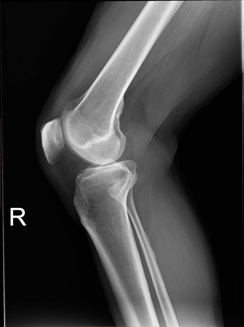
2. Lateral Knee (Mediolateral)
Patient position: Turn onto the affected side. Knee flexed 20-30° (this opens the joint space — a fully extended knee closes it).
Part position: Femoral condyles superimposed (posterior borders aligned), patella perpendicular to the IR.
Central ray: Angled 5-7° cephalad, entering 2.5 cm distal to the medial epicondyle.
Evaluation criteria: Femoral condyles superimposed, patella in profile, open patellofemoral joint space, tibial plateaus not superimposed.
Common Mistake
An under-rotated lateral knee will show the adductor tubercle prominently on the medial femoral condyle. If you see it, the patient is under-rotated — roll them slightly more anterior. Also, too much knee flexion (>30°) closes the joint space.
3. AP Oblique Knee (Internal or External Rotation)
Some protocols include an oblique view. Internal (medial) rotation of 45° opens the proximal tibiofibular joint. External (lateral) rotation of 45° opens the lateral aspect of the knee joint.
Special Projections
Intercondylar Tunnel View (Notch View)
Used to visualize the intercondylar fossa, tibial spines, and posterior femoral condyles. Critical for detecting loose bodies and osteochondral defects.
Patient position: Prone, knee flexed 40-50°. Or supine with the IR against the anterior thigh (Camp-Coventry method).
Central ray: Caudal angle equal to the degree of knee flexion (typically 40° from the horizontal for a 40° flexed knee), centered to the knee joint. In the Holmblad method (kneeling), the CR is perpendicular to the lower leg.
Evaluation criteria: Intercondylar fossa open and well-demonstrated, tibial spines visible, posterior femoral condyles seen.
Sunrise View (Patellar Skyline / Merchant View)
Visualizes the patella and patellofemoral joint. Essential for evaluating patellar tracking, fractures, and degenerative changes.
Patient position: Supine with knee flexed 40-45° over a positioning wedge or sponge. The IR is placed against the anterior thigh.
Central ray: Directed through the patellofemoral joint space, typically with a 30° cephalad angle from the lower legs (or a variable angle depending on knee flexion — the beam should be parallel to the patellofemoral joint).
Evaluation criteria: Patella in profile, patellofemoral joint space open, both femoral condyles symmetric, no superimposition of the tibia on the patella.
| Projection | Knee Flexion | CR Angle | Key Anatomy |
|---|---|---|---|
| AP | 0° (fully extended) | 0° (or 5-7° cephalad) | Femoral/tibial condyles, joint space |
| Lateral | 20-30° | 5-7° cephalad | Patella, patellofemoral joint, femoral condyles |
| Tunnel | 40-50° | 40° caudal (prone) | Intercondylar fossa, tibial spines |
| Sunrise | 40-45° | 30° cephalad | Patella, patellofemoral joint space |
Technical Factors
Typical exposure ranges for an average adult knee (grid, 100 cm SID):
- AP/Lateral: 60-70 kVp, 4-8 mAs (digital CR/DR)
- Tunnel: 65-75 kVp, 6-10 mAs (thicker part, more scatter)
- Sunrise: 55-65 kVp, 3-5 mAs (smaller part, less attenuation)
Always adjust for patient habitus, pathology (osteoporosis drops technique, joint effusion rises it), and AEC if available.
Quick Reference Card
Print this out and keep it in your pocket for clinicals:
- AP: Patella up → center 2 cm below patellar apex → 0° CR
- Lateral: Roll onto affected side → flex 20-30° → condyles superimposed → 5-7° cephalad
- Tunnel: Prone → flex 40-50° → CR 40° caudal
- Sunrise: Supine → flex 40-45° → IR on thigh → CR 30° cephalad