X-Ray Physics Made Simple: kVp, mAs, Density, and Contrast
Why Physics Matters (Even If You Hate It)
Every radiography student asks the same question: "Do I really need to know the physics?" Yes. Because when your image comes out too light and the radiologist sends it back, you need to know whether to increase kVp, mAs, or both — and by how much. Physics isn't just for the registry. It's what makes you a technologist instead of a button-pusher.
The Golden Rule of Radiographic Exposure
kVp controls contrast (and influences density). mAs controls density (quantity of X-rays). Understanding this relationship is critical for radiation safety and dose optimization. (quantity of X-rays). They interact, but understanding which lever does what is the foundation of everything.
The Four Radiographic Qualities
Every X-ray image is evaluated on four properties. If you understand these, you understand physics:
Density (Brightness)
Overall darkness of the image. Controlled primarily by mAs. Higher mAs = more X-ray photons = darker image (more density).
Contrast
Difference between adjacent densities. Controlled primarily by kVp. Lower kVp = higher contrast (more black-and-white). Higher kVp = lower contrast (more shades of gray).
Recorded Detail
Sharpness of structural lines. Controlled by focal spot size, SID, OID, and motion. Smaller focal spot = sharper. Longer SID = sharper.
Distortion
Misrepresentation of true object size or shape. Controlled by alignment (CR, part, IR) and SID/OID relationship. Minimized with proper positioning.
kVp — The Contrast Controller
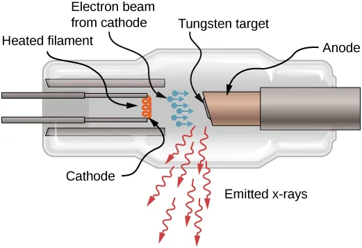
Kilovoltage peak (kVp) determines the energy (quality) of the X-ray beam. Higher kVp = higher-energy photons = more penetration = lower contrast.
The 15% Rule
A 15% increase in kVp approximately doubles the exposure to the IR (same effect as doubling mAs). Conversely, a 15% decrease in kVp approximately halves the exposure.
Clinical Application
If your technique is 70 kVp @ 10 mAs and you want to reduce patient dose by increasing kVp, a 15% increase means: 70 × 1.15 = 80.5 kVp @ 5 mAs (you halved mAs to compensate). The image will have slightly less contrast but the same overall density.
mAs — The Density Controller
Milliampere-seconds (mAs) determines the quantity of X-ray photons produced. It's the product of mA (tube current) and exposure time in seconds.
mAs and density have a directly proportional relationship: double the mAs = double the density (all else being equal). This is the mAs Reciprocity Law.
The Reciprocity Law
100 mA × 0.1 s = 10 mAs produces the same density as 200 mA × 0.05 s = 10 mAs. The product is what matters, not the individual components. This is why you can use a higher mA station with a shorter exposure time to freeze motion — same density, better detail. These principles also apply to CT scanning and radiotherapy. station with a shorter exposure time to freeze motion — same density, better detail.
The Inverse Square Law
The intensity of radiation is inversely proportional to the square of the distance from the source. This is the most frequently tested physics concept on the ARRT exam.
Formula: I₁ / I₂ = (D₂ / D₁)²
Example
If you increase SID from 100 cm to 180 cm, what happens to intensity?
I₁ / I₂ = (180 / 100)² = (1.8)² = 3.24
Intensity drops to 1/3.24 of the original — you'll need ~3.24× more mAs to maintain density.
The Direct Square Law (mAs Compensation)
To compensate for SID changes: mAs₁ / mAs₂ = (SID₁ / SID₂)²
If original technique is 10 mAs at 100 cm SID and you move to 180 cm SID:
10 / mAs₂ = (100/180)² → mAs₂ = 10 × 3.24 = ~32 mAs
Density vs Contrast — The Visual Difference
Here's the clearest way to think about it:
- Too light overall (underexposed): Increase mAs by a factor of 2 (double it)
- Too dark overall (overexposed): Decrease mAs by half
- Too gray (low contrast, "flat" image): Decrease kVp by ~10-15%
- Too black-and-white (high contrast, "chalky"): Increase kVp by ~10-15%
The Grid Factor
Grids improve contrast by absorbing scatter radiation before it reaches the IR. But they also absorb some primary radiation, so you need to increase mAs. The grid conversion factor (Bucky factor) tells you how much:
| Grid Ratio | Bucky Factor | mAs Multiplier |
|---|---|---|
| No grid | 1 | 1× |
| 5:1 | 2 | 2× |
| 8:1 | 4 | 3-4× |
| 12:1 | 5 | 4-5× |
| 16:1 | 6 | 5-6× |
The One-Page Cheat Sheet
Memorize these relationships for the registry and for the control panel:
- mAs ↑ → Density ↑ (directly proportional)
- kVp ↑ → Contrast ↓, Density ↑ (not linear!)
- SID ↑ → Density ↓ (inverse square law)
- Grid ↑ → Contrast ↑, Density ↓ (compensate with mAs)
- Collimation ↑ → Scatter ↓, Contrast ↑ (tighter collimation = better contrast)
- Part thickness ↑ → Density ↓ (more attenuation)
- 15% kVp ≈ Doubling/Halving mAs (for density compensation)